
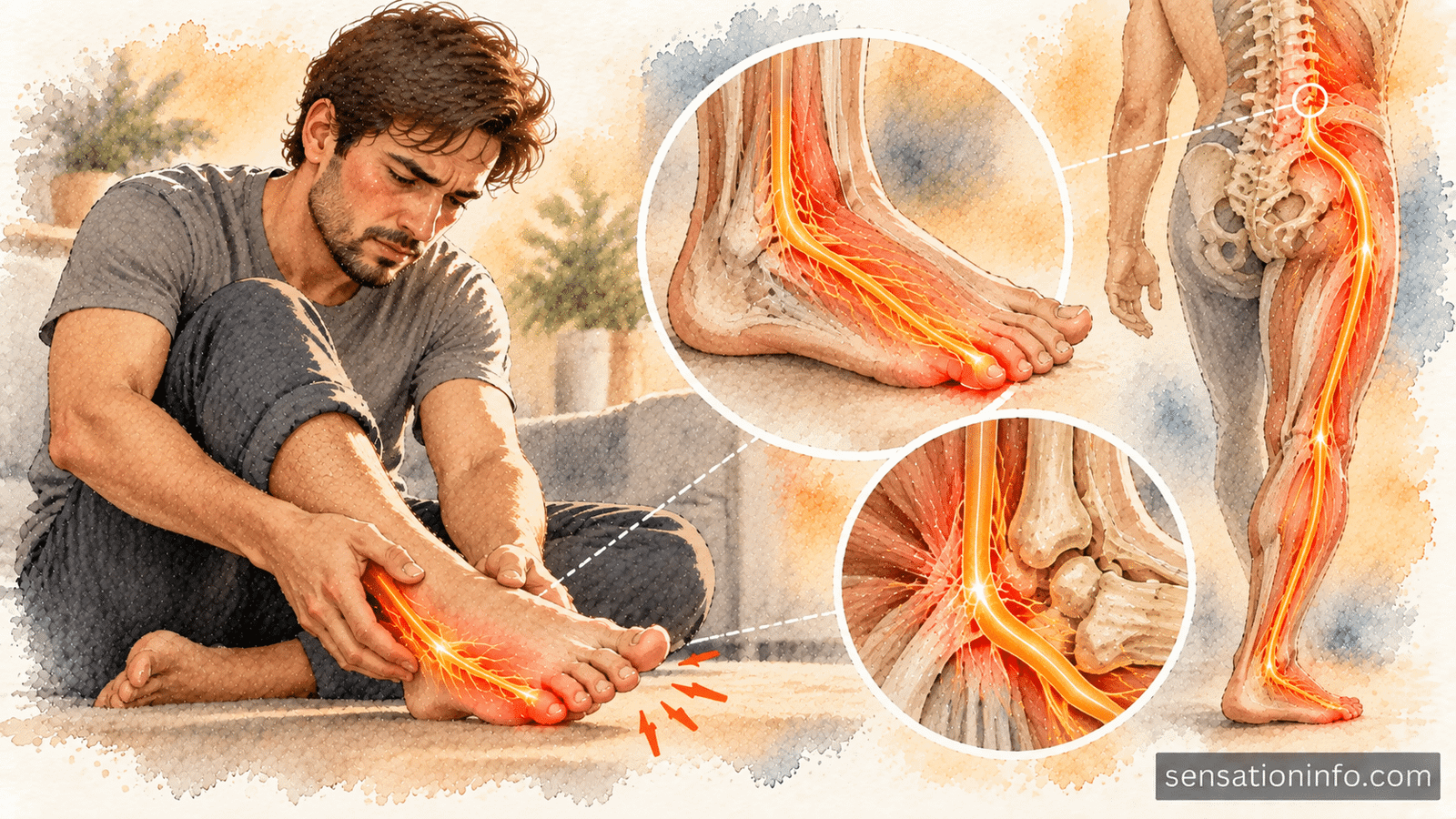

Experiencing numbness in the outer foot and little toe occurs when the sensory pathway running along the lateral border of your foot becomes compressed or irritated. While squeezing into tight footwear or suffering a recent ankle injury can easily pinch the local sural nerve near your heel, this specific sensory loss frequently originates several feet higher in your lower back.
Compression of the S1 nerve root at the L5-S1 spinal level is actually one of the most common culprits, meaning that a structural problem in your lumbar spine can produce uncomfortable sciatic tingling and numbness that bypasses your back and is felt entirely in your foot.
Red Flag Symptoms That Require Immediate Medical Attention
Most cases of outer foot numbness develop gradually from mechanical pressure or nerve irritation. However, certain symptoms indicate severe spinal cord or nerve root compression that requires emergency intervention.
Seek immediate emergency medical care if you experience any of the following warning signs:
- Sudden loss of bowel or bladder control, or difficulty starting urination, which are hallmark signs of cauda equina syndrome
- Progressive numbness in the saddle region, which includes your groin, buttocks, and inner thighs
- Rapidly worsening weakness in both legs, making it impossible to stand, walk, or support your body weight
- Complete inability to push your foot downward against resistance or stand on your tiptoes, known as severe plantarflexion failure
- Numbness that appears suddenly alongside severe trauma, fever, or a personal history of cancer
If your numbness is confined to the outer edge of one foot without saddle numbness or loss of bladder function, you should still arrange a timely consultation with your physician or spine specialist to determine the exact level of nerve compromise.
Understanding the Nerve Pathways to Your Outer Foot
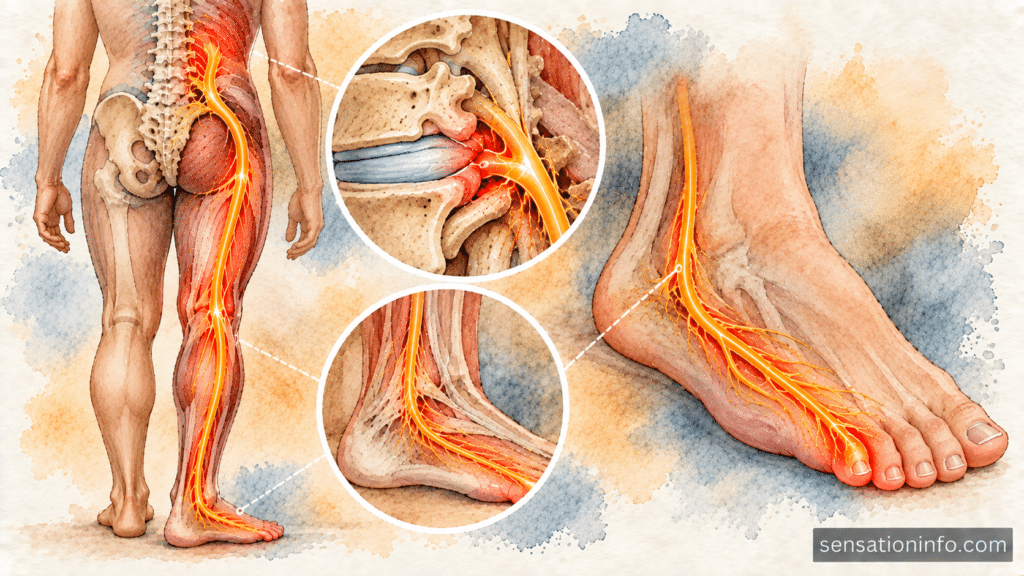
To understand why a lower back issue causes numbness in your smallest toe, it helps to trace the electrical wiring that connects your spine to your foot.
Your nervous system organizes skin sensation into mapped zones known as dermatomes. The S1 dermatome covers the lower back, travels down the back of your thigh and calf, wraps around the outer ankle bone, and finishes along the outer lateral border of the foot, including the fifth toe.
Two primary nerve structures can produce numbness along this specific track:
- The S1 Spinal Nerve Root: Located in your lower back where the fifth lumbar vertebra meets the top of the sacrum, the S1 nerve root exits the spinal column and joins other nerves to form the sciatic nerve.
- The Sural Nerve: Located in your lower leg and ankle, the sural nerve is a peripheral nerve formed from branches of the tibial and common fibular nerves. It carries sensory information directly from the outer ankle, heel, and lateral foot back up to the spinal cord.
Because the sural nerve is composed largely of fibers that originate from the S1 nerve root, a pinched nerve in your lower spine and a pinched nerve at your outer ankle can feel virtually identical in your foot.
Common Causes Originating in the Lower Back
According to clinical data published by the National Institutes of Health, lumbar radiculopathy affects approximately three to five percent of the population over a lifetime, with the S1 nerve root being involved in over forty percent of all lumbosacral disc cases.
L5-S1 Herniated Disc
Intervertebral discs act as shock absorbing cushions between your spinal bones. When the tough outer ring of the disc between the L5 and S1 vertebrae tears, the soft gel interior can push outward directly onto the traversing S1 nerve root. Even a tiny disc bulge at this junction can disrupt nerve signaling, sending radiating numbness and sciatica and leg sensations down your calf and foot.
Lumbar Spinal Stenosis and Foraminal Narrowing
As the spine ages, arthritis, thickened ligaments, and bone spurs can narrow the spinal canal or the neural foramina where nerve roots exit the spine. When the exit window for the S1 nerve root shrinks, the bone spurs physically pinch the nerve fibers during standing or walking, producing gradual numbness in the outer foot that improves when leaning forward or sitting down.
Degenerative Disc Disease and Spondylolisthesis
When the disc space between L5 and S1 collapses or when one vertebra slips forward over the one below it, the spinal alignment changes. This structural instability stretches or compresses the S1 nerve root, leading to persistent lateral foot numbness, particularly during prolonged physical activity or when experiencing a burning sensation in the lower back from nerve inflammation.
Local and Peripheral Causes in the Foot and Ankle
Not all outer foot numbness originates in the spine. Localized trauma, friction, or compression along the path of the sural nerve in your lower leg can also cut off sensation to the fifth toe.
Tight Footwear and Pressure Points
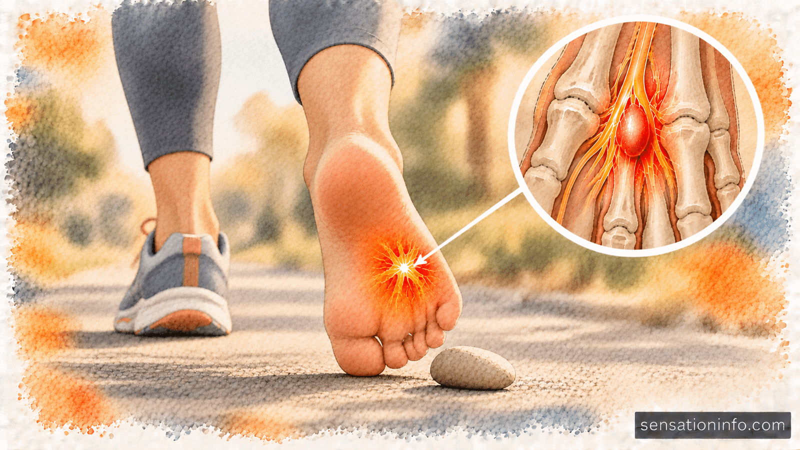
Wearing narrow running shoes, rigid ski boots, or tight work boots can exert excessive physical pressure against the lateral border of your foot and the fifth metatarsal bone. This external compression squashes the terminal branches of the sural nerve, creating temporary numbness, pins and needles, and localized burning in the little toe.
Inversion Ankle Sprains and Fractures
When you roll your ankle outward during a sports injury or fall, the soft tissues on the outer side of your ankle stretch violently. This trauma can stretch, bruise, or tear the sural nerve as it travels behind the lateral malleolus. Similarly, a fracture at the base of the fifth metatarsal bone can damage the nerve endings directly beneath the bone.
Sural Nerve Entrapment
The sural nerve passes through tight fibrous tunnels in the calf and outer ankle. Deep scar tissue from prior surgeries, direct impact to the calf, localized cysts, or chronic swelling can trap the nerve against surrounding fascia, producing isolated sensory loss along the outer foot without any back symptoms. If multiple toes become numb simultaneously, your doctor may also evaluate general numbness in the toes from peripheral nerve disorders to rule out systemic metabolic conditions.
| Diagnostic Feature | S1 Lumbar Radiculopathy (Spine) | Sural Nerve Entrapment (Ankle/Foot) | Tight Footwear Compression |
|---|---|---|---|
| Location of Numbness | Outer foot, little toe, back of calf | Outer border of foot and heel | Little toe and outer foot edge |
| Back or Buttock Pain | Frequent deep aching or sharp shooting | None | None |
| Influence of Posture | Worsens with coughing, sitting, bending | No change with spine movement | Improves immediately when shoes are off |
| Reflex Changes | Diminished or absent Achilles tendon reflex | Normal reflexes | Normal reflexes |
| Motor Weakness | Difficulty standing on tiptoes | None (sural nerve is purely sensory) | None |
What Does Outer Foot and Little Toe Numbness Feel Like?
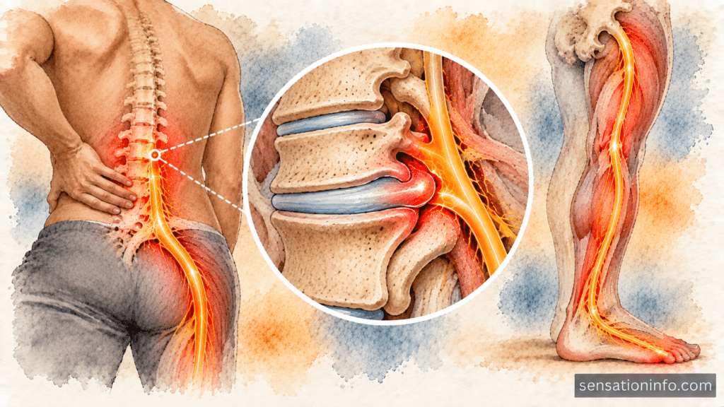
The sensory loss in your outer foot can manifest in several distinct ways depending on the severity of the nerve impingement.
In mild cases, you might feel a sensation similar to wearing a thick sock when your foot is completely bare. Running your fingers across the outside edge of your foot may feel dull, rubbery, or muffled compared to your other foot.
In moderate cases, periods of numbness alternate with uncomfortable buzzing, prickling, or electrical zaps that travel along the outer side of your heel into the small toe. You may notice these sensations intensify when bending forward at the waist or when experiencing tingling in the back and spine provoked by bending.
In severe, chronic nerve compression, the outer foot becomes completely numb to light touch and temperature changes. You may not feel small stones in your shoe, which increases the risk of skin blisters, calluses, or unobserved cuts on the side of your foot.
How Healthcare Providers Diagnose the Source
Accurately determining whether your foot numbness stems from your lower spine or your ankle requires a thorough clinical and electrodiagnostic workup.
Your diagnostic evaluation typically involves:
- Physical and Neurological Examination: Your doctor will test sensation across your foot using a monofilament wire, evaluate your Achilles tendon reflex with a reflex hammer, and assess your calf strength by asking you to perform single-leg heel raises.
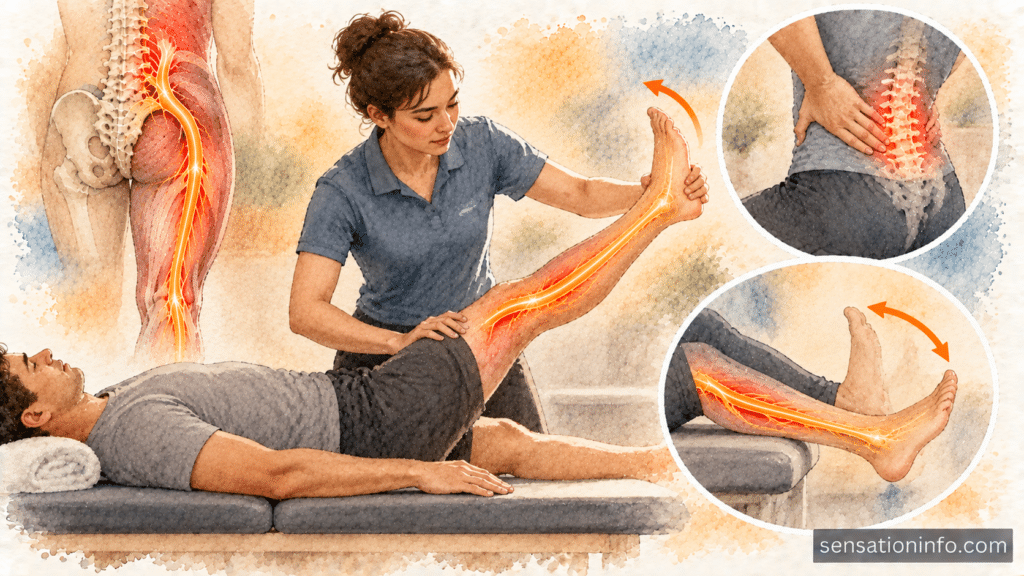
- Straight Leg Raise Test: While you lie on your back, your physician will lift your extended leg. If raising the leg between thirty and seventy degrees reproduces sharp shooting pain or numbness into your foot, the test strongly indicates lumbar disc herniation.
- Lumbar Spine MRI: A high-resolution magnetic resonance imaging scan of your lower back provides detailed cross-sectional views of your L5-S1 disc, revealing whether a disc herniation or bone spur is pressing against the S1 nerve root.
- Nerve Conduction Studies and Electromyography: A nerve conduction study and EMG evaluation measures the speed and strength of electrical signals passing through your leg nerves. This test definitively differentiates between a preganglionic spinal root injury and a postganglionic peripheral sural nerve problem.
- Diagnostic Ankle Ultrasound: If a localized ankle problem is suspected, musculoskeletal ultrasound can visualize scar tissue, neuroma formation, or fluid collections compressing the sural nerve behind your ankle bone.
Medical Treatments for S1 Radiculopathy and Foot Numbness
Treatment focuses on reducing nerve inflammation, relieving mechanical pressure, and restoring normal sensory signaling.
Conservative Medical Care
According to clinical reviews from the Mayo Clinic, approximately seventy to ninety percent of individuals with lumbar radiculopathy improve with conservative management within six to twelve weeks. First-line therapies include:
- Nonsteroidal anti-inflammatory drugs like meloxicam (Mobic) or naproxen (Aleve) to decrease inflammation around the pinched nerve root
- Short courses of oral corticosteroids such as methylprednisolone (Medrol) for rapid reduction of acute nerve root swelling
- Neuropathic pain stabilizers like gabapentin (Neurontin) or pregabalin (Lyrica) to calm abnormal nerve tingling and shooting sensations
Physical Therapy and Spinal Decompression
Dedicated physical therapy is the foundation of long-term recovery for spinal nerve compression. A physical therapist will guide you through:
- Directional preference exercises that encourage disc material to migrate away from the S1 nerve root
- Core stabilization routines to strengthen the deep abdominal and spinal muscles that support the L5-S1 joint
- Neural mobilization and nerve flossing techniques that improve the sliding ability of the sciatic and sural nerves through tight anatomical channels
Lumbar Epidural Steroid Injections
If severe numbness and pain persist despite oral medication and physical therapy, a fluoroscopically guided lumbar epidural steroid injection can deliver concentrated anti-inflammatory medication directly into the epidural space surrounding the inflamed S1 nerve root.
Surgical Decompression
When severe nerve compression causes progressive muscle weakness, unrelenting pain, or signs of cauda equina syndrome, surgical intervention such as a minimally invasive lumbar microdiscectomy may be indicated. The surgeon removes the small fragment of herniated disc pressing on the S1 nerve, immediately relieving mechanical tension.
Practical Home Care and Foot Protection
While undergoing medical treatment, these everyday adjustments can protect your foot and alleviate discomfort:
- Wear wide toe box shoes: Choose footwear that provides ample room along the lateral border of your foot to prevent secondary compression on the sural nerve.
- Avoid prolonged static sitting: Take brief walking breaks every thirty to forty minutes to relieve sustained hydrostatic pressure on your lumbar spinal discs.
- Practice daily foot inspections: Because numb skin cannot feel friction or cuts, inspect the outside edge of your foot and little toe each night for redness, blisters, or skin breaks.
- Use cold and heat therapy properly: Apply an ice pack wrapped in a towel to your lower back for fifteen minutes to reduce acute spinal inflammation. Never apply extreme heat or ice directly to your numb foot, as reduced skin sensitivity increases the risk of thermal burns.
- Maintain neutral sleeping posture: Sleep on your side with a pillow between your knees, or on your back with a pillow under your knees, to keep your lower back in a neutral, decompressed position.
Common Myths About Foot Numbness
- Myth: If your foot is numb, the problem must be located in your foot. Fact: Sensory nerves run continuously from your brain to your toes. A nerve pinched in your lower back will frequently cause symptoms that are felt only at the end of the line in your foot.
- Myth: You always need surgery for a herniated disc causing foot numbness. Fact: The vast majority of lumbar disc herniations shrink and heal naturally through conservative care, physical therapy, and targeted exercises over several weeks to months.
- Myth: Resting in bed for several weeks is the best way to heal a pinched spinal nerve. Fact: Prolonged bed rest weakens stabilizing core muscles and stiffens spinal joints. Controlled, gentle walking and physical therapy promote faster nerve healing and better blood flow.
Prevention Guidelines for Lower Back and Foot Health
Protecting your lumbar spine and peripheral nerves involves proactive daily habits:
- Use proper lifting mechanics by bending your knees, keeping heavy objects close to your chest, and avoiding simultaneous lifting and twisting motions.
- Strengthen your abdominal wall, gluteal muscles, and back extensors through low impact exercises such as swimming, walking, and core stabilization training.
- Optimize your workstation ergonomics with supportive lumbar seating, positioning your hips slightly higher than your knees.
- Replace worn athletic shoes and avoid excessively tight footwear that pinches your fifth metatarsal bone.
- Maintain a healthy body weight to minimize chronic compressive forces across your lower lumbar spinal discs.
Recovery Outlook and Prognosis
The long-term outlook for outer foot and little toe numbness depends on the duration and extent of the nerve compression.
When nerve compression is identified early and treated with conservative physical therapy, most patients notice sensory recovery within six to twelve weeks. Tingling and burning sensations often resolve first, followed by the gradual return of fine touch sensation to the skin.
If nerve compression has been present for many months or years, the microscopic sensory fibers take longer to regenerate. In some long-standing cases, mild residual numbness may persist even after spinal pressure is completely removed, but dedicated physical therapy and core conditioning prevent symptoms from worsening and protect overall mobility.
Key Takeaways
- Numbness in the outer foot and little toe often stems from compression of the S1 nerve root in the lower back rather than an issue in the foot itself.
- The S1 dermatome supplies sensation to the back of the calf, outer ankle, lateral border of the foot, and the fifth toe via the sural nerve.
- Common spinal causes include L5-S1 herniated discs, lumbar spinal stenosis, and bone spurs, while local causes include tight shoes and ankle sprains.
- Loss of bowel or bladder control, progressive saddle numbness, or rapid leg weakness are medical emergencies requiring immediate hospital evaluation.
- Most individuals recover successfully through conservative treatments including physical therapy, anti-inflammatory medications, core stabilization, and ergonomic adjustments.
Frequently Asked Questions
A severe inversion ankle sprain can stretch or scar the sural nerve as it passes behind the outer ankle bone, leading to localized numbness that typically improves over several months with proper rehabilitation.
The straight leg raise test is a physical examination technique where your doctor lifts your straight leg while you lie flat on your back to determine if stretching the sciatic nerve reproduces shooting pain, tingling, or numbness down into your foot.
Surgery is typically considered only if severe numbness and pain do not improve after several weeks of conservative physical therapy and medication, or immediately if you develop progressive muscle weakness or loss of bowel or bladder control.
Doctor’s Recommendation
When you experience persistent numbness in your outer foot that does not improve after changing your shoes or resting, seeking a comprehensive evaluation from a spine or sciatica specialist is critical. An experienced spine physician can perform thorough neurological testing, order targeted spinal imaging, and design an individualized rehabilitation plan to decompress your S1 nerve root before permanent sensory loss develops.
To find highly qualified medical experts who diagnose and treat lumbar disc herniations, sciatica, and nerve compression across the United States, explore the sciatica specialists directory for top physician listings to connect with leading providers in your area.
References
- National Institutes of Health. Lumbosacral Radiculopathy: Clinical Anatomy, Pathophysiology, and Diagnostic Evaluation. National Library of Medicine PubMed Central. 2023.
- Mayo Clinic. Herniated Disk: Symptoms, Causes, Diagnosis, and Treatment Options. Mayo Foundation for Medical Education and Research. 2024.
- Cleveland Clinic. Sciatica: Causes, Symptoms, Diagnosis, and Management. Cleveland Clinic Health Library. 2023.
- American Academy of Orthopaedic Surgeons. Lumbar Spinal Stenosis and Nerve Compression Disorders. OrthoInfo Clinical Patient Education. 2023.


2 Responses